CBCT Scan

What is a CBCT scan used for?
A Cone Beam Computed Tomography (CBCT) scan is widely used in dental and maxillofacial fields to capture detailed 3D images of the teeth, jaw, and surrounding structures. This imaging technique is crucial for planning dental implants, as it accurately assesses bone structure and density, ensuring proper placement. In orthodontics, CBCT scans aid in the precise alignment of braces and tracking tooth movement. They are also essential in endodontics for diagnosing complex root canal issues and in evaluating temporomandibular joint (TMJ) disorders by providing clear images of the joint’s anatomy. Additionally, CBCT is valuable in maxillofacial surgery for detailed pre-operative planning, helping surgeons visualize critical structures before procedures. The scan’s ability to deliver high-resolution images with relatively low radiation exposure makes it an ideal tool for procedures that require meticulous anatomical visualization and precise treatment planning.

Are CBCT scans safe?
CBCT scans are generally considered safe, particularly when compared to traditional CT scans. They use a cone-shaped X-ray beam that emits a lower dose of radiation, reducing the patient’s exposure while still providing high-quality, detailed images. This makes CBCT scans a safer option for repeated imaging, which is often necessary in dental and maxillofacial care. Despite the lower radiation levels, it’s still important to use CBCT scans judiciously, especially in vulnerable populations like children and pregnant women, where minimizing radiation exposure is crucial. The benefits of CBCT scans, such as their ability to provide precise 3D images for accurate diagnosis and treatment planning, often outweigh the risks associated with radiation. However, as with any medical procedure involving radiation, the decision to use CBCT should be based on clinical necessity, with appropriate measures taken to protect the patient and minimize exposure.

What is a CBCT scan?
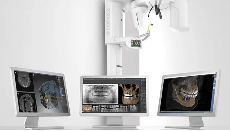
A Cone Beam Computed Tomography (CBCT) scan is a specialized imaging technique that produces detailed three-dimensional images, primarily used in dental and maxillofacial fields. Unlike traditional CT scans, which create images slice by slice, a CBCT scan uses a cone-shaped X-ray beam to capture the entire area in a single rotation, offering a comprehensive view of teeth, bones, soft tissues, and nerve pathways. This technology is vital for precise diagnosis and treatment planning in procedures like dental implants, orthodontics, and TMJ analysis. CBCT scans emit lower radiation compared to traditional CT scans, making them a safer option for patients, especially when multiple scans are necessary. The process is quick, usually completed in under a minute, and is non-invasive, requiring no special preparation. Due to its high-resolution imaging and reduced radiation exposure, CBCT is an essential tool in modern dental and medical practice.

What happens during a CBCT scan?
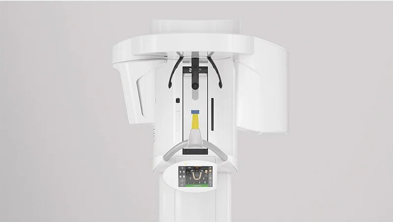
During a Cone Beam Computed Tomography (CBCT) scan, the patient is seated or lies down, depending on the specific equipment used. The technician positions the patient’s head within the scanning machine, ensuring that they remain still to capture accurate images. Once in position, the CBCT machine rotates around the patient’s head in a single, 360-degree motion, using a cone-shaped X-ray beam to capture multiple images from different angles. This process typically takes less than a minute. The images are then compiled by computer software to create a detailed 3D representation of the area being examined, such as the teeth, jaw, or facial bones. The scan is non-invasive and painless, with no special preparation required. The patient is exposed to a relatively low dose of radiation compared to traditional CT scans, making CBCT a safer option for diagnostic imaging and treatment planning in dental and maxillofacial care.


